

Coronavirus disease 2019 (COVID-19), which is zoonotic in origin has caused a great global threat to public health.The World Health Organization (WHO) has declared COVID-19 disease as a pandemic. It most commonly spread through respiratory droplets or aerosol transmission,which urgently demands for better understanding of COVID-19 histopathogenesis.
It wasoriginated in the seafood market of Wuhan, Hubei, China, in early December 2019 with clinical presentations similar to viral pneumonia.
Coronaviruses such as SARS‑CoV2, MERS‑CoV, and SARS‑CoV1 can cause significant morbidity and mortality in infected persons. The severity increases with presence of comorbidities like hypertension, chronic kidney disease, obstructive sleep apnoea and metabolic diseases like diabetes and obesity,11 12 and the pathological changes may even vary between right and left lung
Lung is the most common site of infection for all the three of these viruses, patients presenting symptoms related to the respiratory system such as sore throat, fever, malaise, and respiratory distress, and in worst cases may proceed to respiratory failure. Pulmonary involvement is also responsible for the high viral transmission.
CHANGES IN THE LUNGS:
Interstitial inflammation, diffuse alveolar damage, and necrotizing bronchitis/ bronchiolitis are general histopathological findings of lung in respiratory viral infections. Diffuse alveolar damage is the most commonly observed finding with respiratory virus infections both in acute and late (organizing) stages.
The characteristic features of acute diffuse alveolar damage are intra‑alveolar oedema. Fibrin deposition and formation of hyaline membranes lining the alveolar walls follow. Late diffuse alveolar damage stages are Type II pneumocyte proliferation, granulation tissue formation, followed by collagen deposition.
MICROSCOPIC CHANGES IN THE LUNGS:
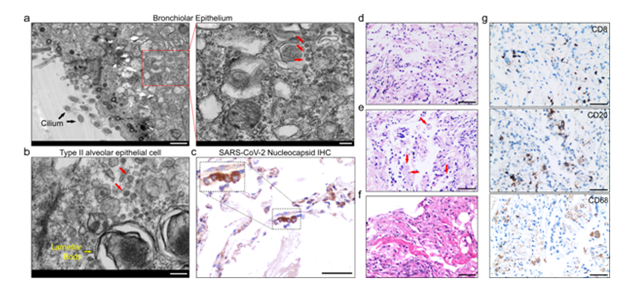
Alveoli: Damaged or atypical enlarged pneumocytes with large nuclei, type II pneumocyte hyperplasia, diffuse alveolar damage (DAD), focal sloughing, hyaline membrane formation, intra-alveolar haemorrhage, intra-alveolar neutrophil infiltration, amphophilic granular cytoplasm and prominent nucleoli characteristic of viral cytopathic-like changes.
Vessels: Oedematous and congested vessels, plug formation, fibrinoid necrosis of the small vasculature, hyaline thrombi in microvessels. Significant deposits of complements—C5b-9 (membrane attack complex), C4d, and mannose binding lectin (MBL)-associated serine protease (MASP)-2, in the microvasculature.
Cellular components: Presence of syncytial giant cells, focal infiltration of immune and inflammatory (lymphocytes and monocytes) and increased stromal cells.
Ultrastructural changes: Viral particles in bronchial mucosal epithelia and type II alveolar epithelia.
Abnormal lung function (ie, restrictive abnormalities, reduced diffusion capacity, and small airways obstruction) has also been identified later on.
These lung function abnormalities appear to be more common among patients with high levels of inflammatory markers, and are often accompanied by evidence of pulmonary fibrosis including interstitial thickening, coarse reticular patterns, and parenchymal bands.Pulmonary fibrosis is associated with permanent pulmonary architectural distortion and irreversible lung dysfunction. Available clinical, radiographic, and autopsy data has indicated that pulmonary fibrosis is central to severe acute respiratory distress syndrome (SARS).
CONCLUSION:
We conclude that, from the available literature, the predictors of pulmonary fibrosis in COVID-19 infection are advanced age, illness severity, length of ICU stay and mechanical ventilation, smoking and chronic alcoholism. With no proven effective targeted therapy against pulmonary fibrosis, risk reduction measures should be directed at limiting the severity of the disease and protecting the lungs from other incidental injuries.
For more info,visit: https://www.santosh.ac.in/
