

The Sectional Anatomy Study Tool is a web-based program to help the user learn human anatomy through images produced by CT-scan (computed tomography) and MRI (magnetic resonance imaging). The user can easily navigate through the stacks of image “slices” of each body section. The label “on” / “off” feature provides users an opportunity for self-assessment.
This tool contains a comprehensive set of both CT and MRI images of the human body in all three planes (i.e., the axial, coronal and sagittal planes).
Areas of Application
Potential users would be individuals who desire to have a thorough knowledge of human anatomy or who need to interpret diagnostic images of the human body typically found in the healthcare setting.
This can include
- Imaging professionals
- Radiologic technologists with specialties in CT (CAT-scan) and MRI (magnetic resonance imaging)
- Students / technologists seeking credentialing in the specialty areas of CT, MRI, PET/CT (positron emission technology / CAT-scan)
- Radiation therapy technologists / students
- Pre-medical and medical students
- Anatomy Students
Advantages of this Program
- Easy to use, intuitive
- Compatible with PCs or Macs
- View using the computer browser
- Images are available in the 3 major body planes
- Image stacks allow students to move forward or backward within the image set, permitting students to develop a 3-dimensional model of the body within their mind
- Labels may be turned on or off for study purposes or self-testing
- Choice of program navigation: mouse, keyboard or both
- Icons demonstrate the level of the image being viewed within the body
- One can quickly and easily jump to another image within the stack of images being viewed
HOW TO WORK ON IT
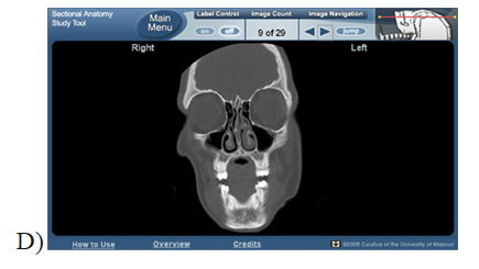
a)The Main Menu page asks you to “Pick A Region” (head, neck, abdomen, etc.) which immediately prompts the user to “Pick A View” (CT and/or MRI image sets) from a drop down menu.
b) The selected image sets demonstrate sequential “slices” through the selected body portion.
c) Navigation tools at the top of the page include: Main Menu button, Label Control (on/off), Image Count, Image Navigation, and an interactive icon that clearly shows the level of the image being viewed within the body region
A BRIEF DEMONSTRATION:
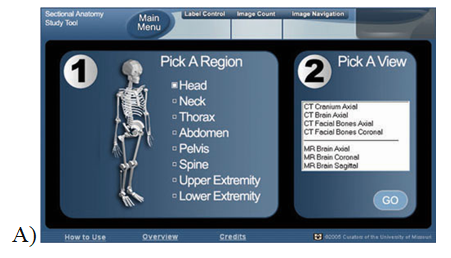
A)Entry Page
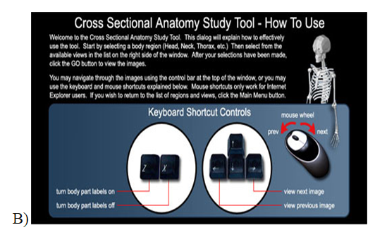
B)How to Use” information screen
C)To select your viewing options:
1. Pick A Region: “Head” selected
2. Pick A View: “CT Facial Bones Coronal” selected
3. Click on “Go”
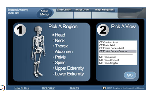
D)Results: CT Facial Bones, coronal view. Page opens with labels off. Note that we have navigated to image 9 of 29. The icon in the upper right corner indicates the approximate level within the body section.
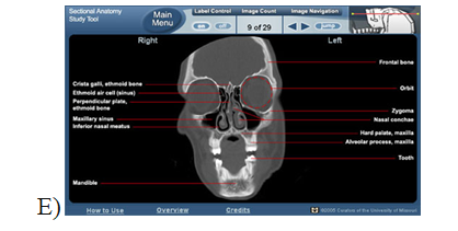
E)Same image with labels turned “on” (on/off switch located on Navigation bar at the top).
